


stog-millets
Bacteria - pearl millet

Contributors to this page: ICRISAT, Patancheru, India (RP Thakur, AG Girish, VP Rao).
|
Contents: |
Brown Spot; Bacterial brown spot
Scientific name
Pseudomonas syringae pv. syringae van Hall.
Other scientific names
Pseudomonas syringae pv japonica
Importance
High
Significance
The diseases caused by this bacterium are very important throughout the world. For instance, bacterial brown spot occurs wherever beans are grown, and canker diseases of fruit trees caused by P. syringae pv. syringae are widespread and may be devastating, causing economical losses.
Symptoms
The symptoms first appear on the lower sides of the leaves as small, water-soaked spots. The spots enlarge, coalesce, and form larger areas that later become necrotic. The bacteria also enter the vascular tissues of the leaf and spread into the stem. The infected area, which is surrounded by a narrow zone of bright, lemon yellow tissue, turns brown, becomes rapidly necrotic, and through coalescence of several small spots, may produce large dead areas of various shapes. The disease produces identical symptoms on the stems, pods and seeds. In addition, light-cream or silver bacterial exudates are often produced on the lesions under moist conditions.
Hosts
Multilateral hosts like gramineae and leguminosae.
Geographic distribution
Worldwide
Biology and transmission
P. syringae pv. syringae is a rod shaped, gram-negative with polar flagella. P. syringae tests negative for arginine dihydrolase and oxidase activity, and forms the polymer levan on sucrose nutrient agar. It is known to secrete the lipodepsinonapeptide plant toxin syringomycin, and it owes its yellow fluorescent appearance when cultured in vitro on King's B medium to production of the siderophore pyoverdin . Pathogen is seed borne and it will spread through the infected seed from season to season (Gaudet and Kokko 1986; Venette et al. 1987). Diseases by P. syringae are favoured by wet, cool conditions - optimum temperature for disease development is around 12 - 25°C, although this can vary according to the pathovar involved. The pathogen is seedborne, and is dispersed between plants via rain splash (Hirano and Upper 1990).
Detection/indexing methods at ICRISAT
- Pre export field inspection and agar plate method.
Treatment/control
- No suitable seed treatment to eradicate infection.
Procedures followed in CGIAR in case of positive test at ICRISAT
- So far not detected at ICRISAT.
References and further reading
Gaudet DA, Kokko EG. 1986. Seedling disease of sorghum grown in southern Alberta caused by seedborne Pseudomonas syringae pv. syringae. Canadian Journal of Plant Pathology 8: 208-217.
Hirano SS, Upper CD. 1990. Population biology and epidemiology of Pseudomonas syringae Annual Reviews in Phytopathology 28:155-177
Venette JR, Lampa RS, Albaugh DA, Nayes JB. 1987. Presumptive procedure (dome test) for detection of seed borne bacterial pathogens in dry beans. Plant Disease 71: 984-990.
Scientific name
Xanthomonas vasicola pv. holcicola (Elliott).
Other scientific names
Bacterium holcicola, Phytomonas holcicola, Pseudomonas holcicola, Xanthomonas campestris pv. holcicola, Xanthomonas holcicola.
Importance
High
Significance
The disease is important only occasionally during springtime under warm weather conditions and becomes less serious during hot and dry summer months.
Symptoms
First symptoms are narrow, water-soaked, transparent leaf streaks, 2-3 mm wide ´ 2-15 mm long, appearing as early as the second leaf stage of the seedling. Lesions soon turn red, become opaque, and at intervals may broaden into somewhat irregularly shaped oval spots with tan centers and narrow red margins. In severe cases, these coalesce to form long irregular streaks and blotches extending across all or much of the leaf blade, with dead tissue bordered by narrow, dark margins between the reddish-brown streaks. Abundant bacterial exudates are produced as light-yellow droplets, which dry to thin white or cream scales (Williams et al. 1978).
Hosts
Panicum miliaceum (broomcorn millet), Setaria italica (foxtail millet), Sorghum halepense (aleppo grass), Sorghum sudanense (Sudan grass), Sorghum bicolor (common sorghum) and Zea mays (maize).
Geographic distribution
Xanthomonas vasicola pv. holcicola is prevalent worldwide (Elliott 1930).
Biology and transmission
Xanthomonas vasicola pv. holcicola is bothsoilborne and seedborne, and is also transmitted by infected plant debris.
Detection/indexing methods at ICRISAT
- Pre export field inspection
Treatment/control
- No seed treatment.
Procedures followed in case of positive test at ICRISAT
- So far not detected.
References and further reading
Elliott C. 1930. Bacterial streak disease of sorghum. Journal of Agricultural Research 40: 963-976.
Williams RJ, Frederiksen RA and Girard JC. 1978. Sorghum and Pearl Millet Disease Identification Handbook. Information Bulletin No. 2. ICRISAT, Patancheru 502324, AP, India. 88pp
Scientific name
Xanthomonas campestris (Pammel) Dowson
Other scientific names
Xanthomonas rubrisorghi, Xanthomonas pennisiti, Xanthomonas annamalaiensis.
Importance
High
Significance
Bacterial leaf streak is of minor importance.
Symptoms
Initial symptoms include narrow, water-soaked, transparent leaf streaks, 2-3 mm wide by 2-15 mm long generally appear from the second leaf stage of the seedlings. Lesions soon turn red, become opaque, and at intervals may broaden into somewhat irregularly shaped oval spots with tan centers and narrow red margins. In severe form, these coalesce to form long irregular streaks extending across the leaf blade. Abundant bacterial exudates are produced as light-yellow droplets, which dry to thin white or cream scales (Williams et al. 1978).
Hosts
Panicum miliaceum (broomcorn millet), Setaria italica (foxtail millet), Sorghum halepense (aleppo grass), Sorghum sudanense (Sudan grass), Sorghum bicolor (common sorghum) and Zea mays (Qhobela and Claflin 1988).
Geographic distribution
It is reported from Nigeria and Senegal.
Biology and transmission
Bacterial colonies are yellow and mucoid on the nutrient agar medium. Bacterial cells are aerobic, motile, gram-negative, rod-shaped which differ pathologically, serologically, and by membrane protein patterns from other pathovars of X. campestris. Cells measure 0.45 ´ 2.25 mm and have one polar flagellum. Optimal growth occurs between 26 and 30 oC (Rangaswami et al. 1961).
Detection/indexing methods at ICRISAT
- Pre export field inspection and agar plate method are used.
Treatment/control
- No seed treatment is available.
Procedures followed in case of positive test at ICRISAT
- So far not detected.
References and further reading
Qhobela M, Claflin LE. 1988. Characterization of Xanthomonas campestris pv. pennamericanum pv. nov., causal agent of bacterial leaf streak of pearl millet. International Journal of Systematic Bacteriology 38: 362-366.
Rangaswami G, Prasad NN, Eswaran KSS. 1961. Two new bacterial diseases of sorghum. Andhra Agricultural Journal 8: 269-272.
Williams RJ, Frederiksen RA, Girard JC. 1978. Sorghum and Pearl Millet Disease Identification Handbook. Information Bulletin No. 2. ICRISAT, Patancheru 502324, AP, India. 88pp.
Fungi - pearl millet
Contributors to this page: ICRISAT, Patancheru, India (RP Thakur, AG Girish, VP Rao).
|
Contents: |
Downy mildew; Green ear disease
Scientific name
Sclerospora graminicola (Sacc.) Schroet.
Other scientific names
Scleropthora macrospora
Importance
Medium
Significance
Yield losses upto 60% were reported from India, and several countries in Africa (Singh et al. 1993).
Symptoms
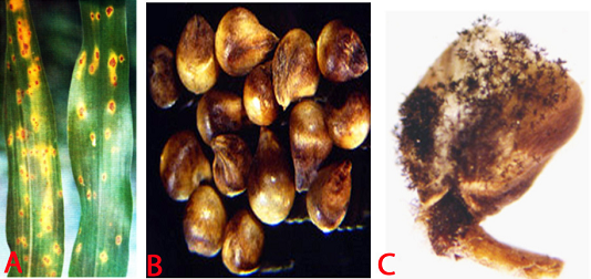
Leaf symptoms begin as chlorosis at the base of the leaf lamina and successive new leaves show a progression of greater leaf coverage. Under conditions of high humidity and moderate temperature, the infected chlorotic leaf area supports a massive asexual sporulation, generally on the abaxial surface of the leaves. Severely infected plants are generally stunted and do not produce panicles. The name 'green ear' stems from the appearance of green panicles due to transformation of floral parts into leafy structures, which can be total or partial (Singh et al. 1993).
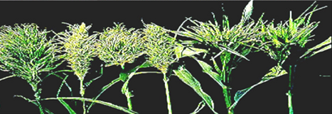 Different malformed green ears (photo: ICRISAT). |
Hosts
S. graminicola is specific to pearl millet.
Geographic distribution
Several countries in Asia and Africa such as Chad, Egypt, Gambia, Malawi, Mozambique, Niger, Nigeria, Zimbabwe, Senegal, South Africa. Also Mali, Burkina Faso, Ivory Coast, Sudan, Kenya, Uganda, Tanzania, Ghana, Togo, Zambia, and a few states in USA.
Biology and transmission
Sclerospora graminicola produces two types of spores, asexual spores known as sporangia, and sexual spores known as oospores. The whitish downy growth of the pathogen on the leaf surface is the “asexual phase”, followed by the “sexual phase” in which oospores are produced within the leaf tissue. Sporangiophores are short, stout, determinate and dichotomously branched structures that emerge from systemically infected leaves through stomata. Sporangia are produced on sterigmata located at the tips of the sporangiophore branches. Sprorangia are hyaline, thin walled, ellipsoid or broadly elliptic and papillate. Sporangia germinate indirectly by producing zoospores. The number of zoospores per sporangium vary from 1-12. Zoospores emerge through a pore produced by the release of the operculum. Zoospores germinate and produce infection in the host. Sexual phase starts with the formation of the oospores in the host. Mature oospores are thick-walled, spherical, and brownish yellow, 22 to 35 um in diameter. Oospores are resting spores and can survive for 8-10 years in field, and cause the primary infection. The pathogen is soilborne and externally seedborne for causing the disease in the succeeding crop season. Secondary spread of the disease occurs through wind borne sporangia (Singh et al. 1993).
Detection/indexing methods At ICRISAT
- Pre export field inspection, and seed washing for oospores detection.
Treatment/control
- Dry seed treatment with metalaxyl @ 2 g a.i. kg-1 seed.
Procedures followed in case of positive test at ICRISAT
- Incineration of infected samples in the field, and rejection of seed samples if oospores are observed under seed washing test.
References and further reading
Ahmed KM, Ravinder Reddy Ch.1993. A Pictorial guide to the identification of seed borne fungi of sorghum, pearl millet, finger millet, chickpea, pigeon pea and groundnut. Information Bulletin No. 34. Patancheru, A.P. 502 324 India: International Crops Research Institute for the semi-Arid Tropics. 200 pp.
Chakrabarty SK, Anitha K, Girish AG, Sarath Babu B, Prasada Rao RDVJ, Varaprasad KS, Khetarpal RK, Thakur RP. 2005. Germplasm exchange and quarantine of ICRISAT mandate crops. Information Bulletin No. 69. Rajendranagar 500 030, Andhra Pradesh, India: National Bureau of Plant Genetic Resources; and Patancheru 502 324, Andhra Pradesh, India: International Crops Research Institute for the Semi Arid Tropics. 80pp.
Singh SD, King SB, Werder J. 1993. Downy mildew disease of pearl millet. Information Bulletin No. 37. Patancheru, AP 502 324, India: International Crops Research Institute for the Semi Arid Tropics. 36pp.
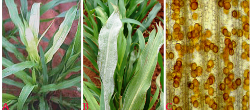 Downy mildew (Sclerospora graminicola) of pearl millet: (A)chlorosis on leaves; (B)sporangial growth on lower surface of leaf and (C)oospores (photos: ICRISAT). |
Scientific name
Claviceps fusiformis Loveless.
Other scientific names
Claviceps microcephala
Importance
High
Significance
The disease assumes special importance because grain is contaminated by grain-replacing sclerotia which contain alkaloids that affect the health of human beings and animals. Losses in grain yield due to this disease have been estimated as high as 58-70% in F1 hybrids (Thakur and King 1988).
Symptoms
Ergot can be identified when creamy to pinkish mucilaginous droplets called ‘honeydew’ ooze from the infected florets on the panicles. These droplets contain numerous asexual conidia. Within 10-15 days these droplets dry out into hard, dark brown to black structures called sclerotia. These are larger than seed and with a pointed apex, which protrude from the florets in place of the grain.
Hosts
Cenchrus ciliaris (buffel grass)and Panicum antidotale (blue panic grass).
Geographic distribution
The disease is distributed in India, Pakisthan, and several countries in Africa including Botswana, Burkina Faso, Gambia, Ghana, Malawi, Niger, Nigeria, Senegal, Somalia, Tanzania, Uganda, Zambia, and Zimbabwe (Thakur and King 1988).
Biology and transmission
Sclerotia are elongated to round in shape, light pink to dark brown/dark in color, hard to brittle with cavities. These germinate by producing 1-17 fleshy, purplish stipes, 6-26 mm long. Each stipe bears at its apex a globular capitulum which is light to dark brown with numerous perithecial projections. Perithecia are pyriform and are embedded in the somatic tissue in the peripheral region of the capitula. Asci are interspersed with paraphyses in the perithecia and emerge through ostioles. These asci are long and hyaline with apical pores. The thread-like ascospores are hyaline and nonseptate. These cause the initial infection in the field. The fungus produces two types of conidia - macro and micro conidia. Macroconidia are hyaline, fusiform, unicellular, and germinate by producing 1-3 germ tubes from their ends or sides. Microconidia are hyaline, globular, unicellular,, and germinate by producing only one germ tube. Both macro- and microconidia are produced on the tips of the germ tubes that are produced in chains. Disease is spread through soil borne sclerotia, sometimes as contaminated seed with sclerotia from season to season. Secondary spread is occurred through wind born conidia and also through rain splashes and insects (Thakur and King 1988).
Detection/indexing methods at ICRISAT
- Pre export field inspection, and seed examination for detection the sclerotia using magnifying lens.
Treatment/control
- Nil
Procedures followed in case of positive test at ICRISAT
- Incineration of infected samples in the field, and rejection of seed samples if found positive in seed examination.
References and further reading
Ahmed KM, Ravinder Reddy Ch.1993. A Pictorial guide to the identification of seed borne fungi of sorghum, pearl millet, finger millet, chickpea, pigeon pea and groundnut. Information Bulletin No. 34. Patancheru, A.P. 502 324 India: International Crops Research Institute for the Semi-Arid Tropics. 200 pp.
Chakrabarty SK, Anitha K, Girish AG, Sarath Babu B, Prasada Rao RDVJ, Varaprasad KS, Khetarpal RK, Thakur RP. 2005. Germplasm exchange and quarantine of ICRISAT mandate crops. Information Bulletin No. 69. Rajendranagar 500 030, Andhra Pradesh, India: National Bureau of Plant Genetic Resources; and Patancheru 502 324, Andhra Pradesh, India: International Crops Research Institute for the Semi Arid Tropics. 80pp.
Thakur RP, King SB. 1988. Ergot disease of pearl millet. Information Bulletin no. 24. Patancheru, AP. 502324. India: International Crops Research Institute for the Semi Arid Tropics. 24pp.
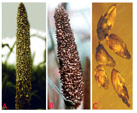 Ergot (Claviceps fusiformis) of pearl millet: (A)honydew stage; (B)sclerotial stage and (C)slerotia (photos: ICRISAT). |
Blast; Leaf blast; Brown leaf spot; Pyricularia leaf spot
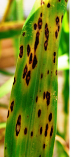 Pyricularia Leaf Spot (photo: ICRISAT) |
Scientific name
Pyricularia grisea (Cke.) Sacc.
Other scientific names
Pyricularia penniseti, Pyricularia setariae.
Importance
Medium
Significance
Leaf blast causes early death of seedlings under humid conditions. It causes the discoloration of the forage and can reduce grain and forage production moderate to heavy .
Symptoms
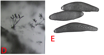
Lesions on foliage are elliptical or diamond-shaped; approximately 2.5-3.5 ´ 1.5-2.5 mm. Lesion centers are grey and water-soaked when fresh but turn brown upon drying. Lesions are often surrounded by a chlorotic halo which will turn necrotic, giving the appearance of concentric rings.
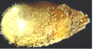 mycelial growth on seed(photo: ICRISAT) |
Hosts
Pennisetum glaucum (pearl millet), Pennisetum purpureum (napier grass).
Geographic distribution
In several countries in the world where warm and humid conditions prevail over a period blast appears on pearl millet.
Biology and transmission
Conidia are pyriform, hyaline, and mostly 3-celled with a small appendage on the base cell. Conidia measure approximately 17.5-30.8 ´ 5.9-8.8 mm (Mehta et al. 1953). Germination, appresoria formation, and invasion of host cells are more at 25°C (Yadava and Agnihotri 1980). Transmission occurs through windborne conidia. It is also reported as seed borne (Singh and Pavgi 1977).
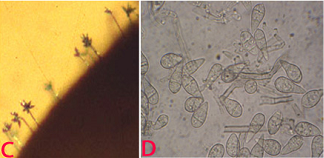 (C)conidiophore with conidia on seed and (D)conidia. (photos: ICRISAT) |
Detection/indexing methods at ICRISAT
- Pre export field inspection and blotter test.
Treatment/control
- Not known
Procedures followed in case of positive test at ICRISAT
- Incineration of infected samples in the field, and rejection of seed samples if found positive in blotter test.
References and further reading
Ahmed KM, Ravinder Reddy Ch.1993. A Pictorial guide to the identification of seed borne fungi of sorghum, pearl millet, finger millet, chickpea, pigeon pea and groundnut. Information Bulletin No. 34. Patancheru, A.P. 502 324 India: International Crops Research Institute for the semi-Arid Tropics. 200 pp.
Chakrabarty SK, Anitha K, Girish AG, Sarath Babu B, Prasada Rao RDVJ, Varaprasad KS, Khetarpal RK, Thakur RP. 2005. Germplasm exchange and quarantine of ICRISAT mandate crops. Information Bulletin No. 69. Rajendranagar 500 030, Andhra Pradesh, India: National Bureau of Plant Genetic Resources; and Patancheru 502 324, Andhra Pradesh, India: International Crops Research Institute for the Semi Arid Tropics. 80pp.
Mehta PR, Singh B, Mathur SC. 1953. A new leaf spot disease of bajra (Pennisetum typhoides Staph and Hubbard) caused by a species of Pyricularia. Indian Phytopathology 5:140-143.
Singh DS, Pavgi MS. 1977. Perpetuation of Pyricularia penniseti causing brown leaf spot of bajra. Indian Phytopathology 30: 242-244.
Yadava RKS, Agnihotri JP. 1980. Epidemiology of Pyricularia leaf spot of bajra. Indian Phytopathology 33:150.
Scientific name
Moesziomyces penicillariae (Bref.) Vanky.
Other scientific names
Tolyposporium penicillariae, Tolyposporium senegalense
Importance
High
Significance
In general, grain loss of 5-20% has been reported (Chahal et al. 1994).
Symptom
In the infected florets, the ovaries are converted into sori. The sori are larger than grains and appear as enlarged, oval to conical bodies projecting beyond the glumes in place of grains. Initially the sori are bright green but later turn brown to black. When the sori mature the membrane rupture and release brown to black mass of spores (Thakur and King 1988).
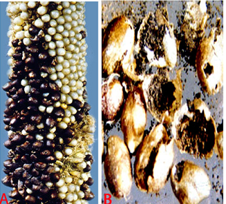 Smut (Moesziomyces penicillariae) of pearl millet: (A)smut sori on panicles (photo: ICRISAT) and (B)sori with teliospores (photo: Chahal et al.1994)/ |
Host
Specific to pearl millet.
Geographic distribution
USA, India, Zimbabwe, Senegal, Chad, Niger, Nigeria, Zambia, Sudan, Cameroon, Burkina Faso, Ghana, Mali, Tanzania (Rachie and Majmudar 1980).
Biology and transmission
The teleutospores occur in compact, ball-like masses called spore balls. Spore balls are circular to near polyhedral. The number of teleutospores aggregated in balls varies from 200 to 1400. Teleutospores are mostly angular to round, light brown in color and 7-12 mm in diameter. Teleutospores germinate sporadically and produce 4 celled promycelium laterally and terminal. Variation in germination patterns of teleutospores occurs while they are held in the spore balls, and the sporidia are produced on branched hyphae in chains (Thakur and King 1988). These sporidia are the main propagules for the cause of the disease. Mostly the teleutospores are wind borne for the secondary spread of the disease. Sometimes the teleutospores are attached to the surface of the seed and act as primary source of inoculum. Teleutospores are also soilborne.
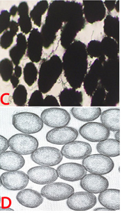 (C)spore masses and (D)spore balls (photos: ICRISAT). |
Detection/indexing methods at ICRISAT
- Pre export field inspection and seed washing test. A new method is developed for elimination of spores from contaminated seed.
Treatment/control
- Spore balls (teleutospores) have been removed by salvaging the smut-contaminated seeds by mixing with sand and ethanol for 3 min while stirring.
Procedures followed in case of positive test at ICRISAT
- Incineration of infected samples in the field, and rejection of seed samples with positive test under seed washing test.
References and further reading
Ahmed KM, Ravinder Reddy Ch.1993. A Pictorial guide to the identification of seed borne fungi of sorghum, pearl millet, finger millet, chickpea, pigeon pea and groundnut. Information Bulletin No. 34. Patancheru, A.P. 502 324 India: International Crops Research Institute for the semi-Arid Tropics. 200 pp.
Chahal SS, Thakur RP, Mathur SB. 1994. Seed borne diseases and seed health testing of pearl millet. Danish Government Institute of Seed Pathology for developing countries, Copenhagen, Denmark. 72pp.
Chakrabarty SK, Anitha K, Girish AG, Sarath Babu B, Prasada Rao RDVJ, Varaprasad KS, Khetarpal RK, Thakur RP. 2005. Germplasm exchange and quarantine of ICRISAT mandate crops. Information Bulletin No. 69. Rajendranagar 500 030, Andhra Pradesh, India: National Bureau of Plant Genetic Resources; and Patancheru 502 324, Andhra Pradesh, India: International Crops Research Institute for the Semi Arid Tropics. 80pp.
Rachie KO, Majmudar JV. 1980. Pearl millet. University Park, Philadelphia, USA: Pennsylvania University press. 307pp.
Thakur RP, King SB. 1988. Smut disease of pearl millet. Information Bulletin No. 25. Patancheru, AP 502 324, India: International Crops Research Institute for the Semi-Arid.
Scientific name
Bipolaris setariae (Saw.) Shoem. [anamorph]
Cochliobolus setariae [Teleomorph]
Other scientific names
Drechslerasetariae
Importance
Medium
Significance
Infection at seedling stage results in death of plants and reduces crop stand in the field (Shetty et al. 1982). Infected plants produce discolored grains and seed of poor quality (Kameswara Rao et al. 2002).
Symptom
Foliar symptoms vary, as brown flecks, fine linear streaks, small oval spots; large irregular oval, oblong, or almost rectangular spots measuring 1-10 ´ 0.5-3 mm. Large fusiform lesions are sometimes produced. Lesions may expand and coalesce. Lesions may be solid dark brown but usually become tan or grayish brown with distinct dark brown border (Luttrell 1954).
 Leaf spot (Bipolaris setariae) of pearl millet: (A)leaf spots (photo: Chahal et al.1994); (B)infected seed and (C)fungal growth on seed (photos: ICRISAT). |
Host
Pennisetum glaucum (pearl millet), Pennisetum purpureum (napier grass), Panicum fasciculatum Swartz (browntop millet), Saccharum officinarum (sugarcane), Zea mays (maize), Sorghum bicolor (sorghum), Paspalum scrobiculatum (little millet), Panicum miliaceum (kodo millet), Hordeum vulgare (barley), Triticum aestivum (wheat), Avena sativa (oat), Imperata cylindrica (Cogongrass) and Cynodon dactylon (bermudagrass).
Geographic distribution
Drechslerasetariae is worldwide in distribution including USA, Hawaii, India, Japan, Zimbabwe and Zambia.
Biology and Transmission
The mycelium is dark and individual hyhae are irregularly branched with rough surfaces.Conidiophores are mostly hypophyllous, simple, 2-8 septate, erect, cylindrical, brown, slightly swollen at the base and geniculate at the apex. They are 72-199 mm in length and 5.6 to 9 mm in width. Conidia are acrogenous, 39-12 mm, 4-10 septate, ellipsoid, straight or slightly curved, pale to moderately dark brown and thin walled (Chahal et al. 1994). The pathogen is both soil borne and seed borne.
 (D)conidiophores with conidia and (E)conidia (photos: ICRISAT). |
Detection/indexing methods used in CGIAR at ICRISAT
- Pre export field infection and blotter test (Girish et al. 2001).
Treatment/control
- Rejection of the seed samples.
Procedures followed in case of positive test at ICRISAT
- Incineration of infected samples in the field, and rejection of seed samples if found positive under blotter test.
References and further reading
Ahmed KM, Ravinder Reddy Ch.1993. A Pictorial guide to the identification of seed borne fungi of sorghum, pearl millet, finger millet, chickpea, pigeon pea and groundnut. Information Bulletin No. 34. Patancheru, A.P. 502 324 India: International Crops Research Institute for the semi-Arid Tropics. 200 pp.
Chahal SS, Thakur RP, Mathur SB. 1994. Seed borne diseases and seed health testing of pearl millet. Danish Government Institute of Seed Pathology for developing countries, Copenhagen, Denmark. 72pp.
Chakrabarty SK, Anitha K, Girish AG, Sarath Babu B, Prasada Rao RDVJ, Varaprasad KS, Khetarpal RK, Thakur RP. 2005. Germplasm exchange and quarantine of ICRISAT mandate crops. Information Bulletin No. 69. Rajendranagar 500 030, Andhra Pradesh, India: National Bureau of Plant Genetic Resources; and Patancheru 502 324, Andhra Pradesh, India: International Crops Research Institute for the Semi Arid Tropics. 80pp.
Girish AG, Singh SD, Chakrabarty SK, Prasada Rao R.D.V.J, Surender A, Varaprasad KS, Bramel PJ. 2001. Seed microflora of five ICRISAT mandate crops. Seed Science and Technology Journal 29: 429-443.
Kameswara Rao N, Bramel Cox P, Reddy KN, Singh SD, Girish AG, Appa Rao S, Mahalakshmi V. 2002. Optimizing seed quality during germplasam regenration in pearl millet. Genetic Resources and Crop Evolution 49: 153-157, Kluwer Academic Publishers, Printed in Netherlands.
Luttrell ES. 1954. Diseases of pearl millet in Georgia. Plant Disease Reporter 38: 507-514.
Shetty HS, Mathir SB, Neergaard P, Safeeula KM. 1982. Drechslera setariae in Indian pearl millet seeds, its seed-borne nature, transmission and significance. Transactions of the British Mycological Society 78: 170-173.
Guidelines for the safe transfer of finger millet germplasm
Contributors to this page: ICRISAT, Patancheru, India (RP Thakur, AG Girish, VP Rao).
Technical Guidelines for the Safe Transfer of Germplasm and the Protection of CGIAR Germplasm Banks
Pathogens of quarantine significance of finger millet tested by the Germplasm Health Laboratory of ICRISAT
Finger millet [Eleusine coracana L.]
|
Bacteria
|
|
Pseudomonas syringae pv. syringae.
|
|
Xanthomonas vasicola pv. holcicola
|
|
Xanthomonas campestris (Pammel) Dowson
|
|
Fungi
|
|
Cochliobolus lunatus
|
|
Insects
|
|
Plodia interpunctella
|
|
Prostephanus truncates
|
|
Tribolium castaneum
|
Insects - pearl millet
Contributors to this page: ICRISAT, Patancheru, India (RP Thakur, AG Girish, VP Rao).
|
Contents: |
Scientific name
Plodia interpunctella Hubner.
Other scientific names
Not known
Importance
High
Significance
Indian meal moth is a common grain-feeding pest. It can infest a variety of products and is perhaps the most economically important insect pest of processed food. Infestations of P. interpunctella can cause direct product loss and indirect economic costs through pest control costs, quality losses, and consumer complaints (Phillips et al. 2000).
Symptoms
Plodia interpunctella is an external feeder. The larvae continuously spin a silken web both inside and on top of the grain surface, and feed within the web. The webbing contains larval excreta (frass) and exuvia (cast skins), and gives an unpleasant odor to the infested commodity. The infested commodity is sometimes covered on the surface with a thick mat of silken webbing (Mohandass et al. 2007).
Biology and transmission
Female moths lay between 60 and 400 eggs (Lyon 1991) on a grain surface, which are ordinarily smaller than 0.5 mm and not sticky. The eggs hatch in 2 to 14 days. The moth larvae are off-white with brown heads. When these larvae mature, they are usually about 12 mm long. The larval stage lasts from 2 to 41 weeks, depending on the temperature. Adult moths are 8–10 mm long with 16–20 mm wingspans. The outer half of their forewings is bronze, copper, or dark gray in color, while the upper half are yellowish-gray, with a dark band at the intersection between the two. The entire life cycle may range from 30 to 300 days (Mohandass et al. 2007).
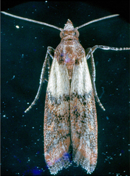 Indian Meal Moth (Plodia interpunctella) of finger millet (photo: ICRISAT). |
Hosts
Arachis hypogaea (groundnut), Oryza sativa (rice), Prunus (stone fruit), stored products (dried stored products), Triticum aestivum (wheat), Zea mays (maize), Avena sativa (oats), Corylus, Helianthus annuus (sunflower), Hordeum vulgare (barley), Juglans regia (Carpathian walnut), Pistacia vera (pistachio), Prunus dulcis (almond) and Theobroma cacao (cocoa).
Geographic distribution
Plodia interpunctella is cosmopolitan in distribution especially in warm climates. In cool temperate countries, it can survive in heated buildings.
Detection/indexing methods at ICRISAT
- The primary detection method is through pheromone-based trapping of males (Phillips et al. 2000). The pheromone commonly referred to as ‘‘ZETA’’ was one of the first commercial pheromones for stored-product insects, and the response of males to this pheromone has been well documented.
Treatment/control
- Fumigation with methyl bromide @ 32 g/m3 for 4 hr followed by seed treatment with chlorpyriphos at 3 g/kg-1 seed (Ghanekar et al. 1996; Toews et al. 2006).
Procedures followed in case of positive test at ICRISAT
- Infested samples are rejected in case of positive test.
References and further reading
Chakrabarty SK, Anitha K, Girish AG, Sarath Babu B, Prasada Rao RDVJ, Varaprasad KS, Khetarpal RK, Thakur RP. 2005. Germplasm exchange and quarantine of ICRISAT mandate crops. Information Bulletin No. 69. Rajendranagar 500 030, Andhra Pradesh, India: National Bureau of Plant Genetic Resources; and Patancheru 502 324, Andhra Pradesh, India: International Crops Research Institute for the Semi Arid Tropics. 80pp.
Ghanekar AM, Ranga Rao GV, Murthy KS, Surender A, Shaik Babu Saheb. 1996. Seed protectants for healthy exports. Indian Journal of Plant Protection 24: 37-43
Lyon WF. 2006. Ohio State University Extension Fact Sheet. Entomology. Indianmeal Moth Ohio State University. Retrieved on 2006-09-07.
Mohandass S, Arthur FH, Zhu KY, Throne JE. 2007. Biology, management of Plodia interpunctella (Lepidoptera: Pyralidae) in stored products. Journal of Stored Products Research 43: 302-311.
Phillips TW, Berbert RC, Cuperus G.W. 2000. Post-harvest integrated pest management. In: Francis, F.J. (Ed.), Encyclopedia of Food Science and Technology. 2nd ed. Wiley Inc., New York, pp. 2690–2701.
Toews MD, Campbell JF, Arthur FH. 2006. Temporal dynamics and response to fogging or fumigation of stored-product Coleoptera in a grain processing facility. Journal of Stored Products Research 42:480-498.
Scientific name
Sitotroga cerealella Olivier.
Other scientific names
Tinea hordei
Importance
High
Significance
Grain losses due to grain moth infestation have been recorded up to 50%. High temperature and lack of storage hygiene are the major factors resulting in insect infestation (Seifelnasr 1992).
Symptoms
Infestation can begin in the fields. In storage, the infestation is confined to the upper layer of the grains. The larva bores into the grain and remains there until it emerges as an adult from round emergence holes. The infested grain is completely hallowed out and filled with larval excreta.
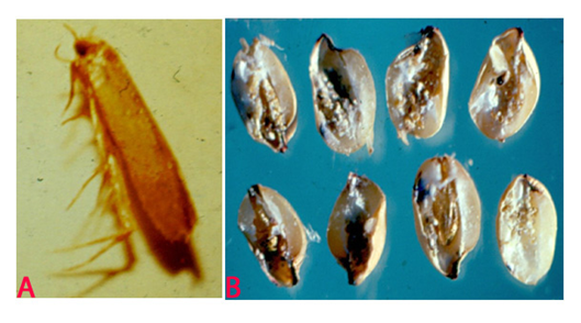 Angoumois Grain Moth (Sitotroga cerealella) of pearl millet: (A)adult and (B)infested grains (photos: ICRISAT). |
Hosts
Avena sativa (oats), Hordeum vulgare (barley), Oryza sativa (rice), Sorghum bicolor (Sorghum), Secale cereale (rye), Triticum aestivum (wheat), Triticum spelta (spelt) and Zea mays (maize).
Geographic distribution
It is cosmopolitan in distribution. It is one of the primary pests of the stored cereals grain, but is also known to attack maize, wheat, and barley in the storage godowns.
Biology
The female can lay up to 400 eggs that are deposited indiscriminately on or between the grains, on the panicles in the field, or in the storage. The egg is white and oval, but soon turns bright red and hatches within a week. The tiny larva crawls about searching for a comparatively weak spot through which it enters the grain, and feeds on the internal contents. The larval period is 2-3 weeks. The moth emerges within a week (Teetes et al. 1983).
Detection/indexing methods at ICRISAT
- Dry seed examination
Treatment/control
- Fumigation with methyl bromide by 32 g/m3 for 4 hr followed by seed treatment with chlorpyriphos at 3 g/kg-1 seed (Ghanekar et al. 1996).
Procedures followed in case of positive test at ICRISAT
- Highly infested samples are rejected
References and further reading
Chakrabarty SK, Anitha K, Girish AG, Sarath Babu B, Prasada Rao RDVJ, Varaprasad KS, Khetarpal RK, Thakur RP. 2005. Germplasm exchange and quarantine of ICRISAT mandate crops. Information Bulletin No. 69. Rajendranagar 500 030, Andhra Pradesh, India: National Bureau of Plant Genetic Resources; and Patancheru 502 324, Andhra Pradesh, India: International Crops Research Institute for the Semi Arid Tropics. 80pp.
Ghanekar AM, Ranga Rao GV, Murthy KS, Surender A, Shaik Babu Saheb. 1996. Seed protectants for healthy exports. Indian Journal of Plant Protection 24: 37-43.
Seifelnasr YE.1992. Farmers’ perceptions on management practices of insect pests on stored sorghum in southwestern Ethiopia. Crop Protection 26: 1817-1825.
Teetes GL, Seshu Reddy KV, Leuschner K, House LR. 1983. Sorghum insect identification handbook. Information bulletin No.12. Patancheru, A.P. , India: International Crops Research Institute for Semi-Arid Tropics. 125pp.
Scientific name
Prostephanus truncatus Horn.
Other scientific names
Dinoderus truncatus
Importance
High
Significance
The grain damage by borer ranges from 19 to 38% and the mean dry weight loss between 5% and 20%. The quantity of grain dust produced weighed between 6 to 29 g kg-1 grains (Shires 1977; Mailafiya et al. 2008).
Symptoms
Adults attack stored seed/grain with intact sheaths by boring into the base of seed making neat round holes. As they tunnel from grain to grain large quantities of grain dust is generated. Adult females lay eggs in chambers bored at right angles to the main tunnels.
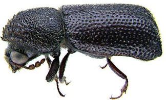 Larger Grain Borer (Prostephanus truncatus) of finger millet (photo: www.agrsci.dk/). |
Hosts
Manihot esculenta (cassava), stored products (dried stored products), Zea mays (maize), Dioscorea (yam) and Triticum aestivum (wheat).
Geographic distribution
Prostephanus truncatus is indigenous in Central America, tropical South America, and the extreme south of the USA as a major pest. It is also distributed in Africa and Europe, and has restricted distribution in India (Farrell 2000).
Biology
The adult has the typical cylindrical bostrichid shape with body length of 3-4.5 mm. The declivity is flattened and steep, and has many small tubercles over its surface. The limits of the declivity, apically and laterally, are marked by a carina. The antennae are 10-segmented and have a loose three-segmented club; the 'stem' of the antenna is slender and clothed with long hairs and the apical club segment is as wide as, or wider than, the preceding segments. The larvae are white, fleshy and sparsely covered with hairs. They are parallel-sided, i.e. they do not taper. The legs are short and the head capsule is small relative to the size of the body (Farrell 2000).
Detection/indexing methods at ICRISAT
- Not detected.
Treatment/control
- Not suggested due to non-detection of the pest.
Procedures followed in case of positive test at iCRISAT
- Infested samples are rejected
References and further reading
Chakrabarty SK, Anitha K, Girish AG, Sarath Babu B, Prasada Rao RDVJ, Varaprasad KS, Khetarpal RK, Thakur RP. 2005. Germplasm exchange and quarantine of ICRISAT mandate crops. Information Bulletin No. 69. Rajendranagar 500 030, Andhra Pradesh, India: National Bureau of Plant Genetic Resources; and Patancheru 502 324, Andhra Pradesh, India: International Crops Research Institute for the Semi Arid Tropics. 80pp.
Farrell G. 2000. Dispersal, phenology and predicted abundance of the larger grain borer in different environments. African Crop Science Journal 8: 337–43.
Mailafiya DM, Ayertey JN, Cudjoe AR. 2008.Damage and weight loss potential of Prostephanus truncates Horn (Coleoptera: Bostrichidae) on sorghum grain: implication to cereal grain storage in sub-Saharan Africa. International Journal of pure and applied sciences 2: 28-35.
Shires SW. 1977. The ability of Prostephanus truncatus (Horn) to damage and breed on several food commodities. Journal of Stored Products Research13: 205-208.
Scientific name
Tribolium castaneum Herbst.
Other scientific names
Not known
Importance
High
Significance
Important in stored pearl millet grain in several countries.
Symptoms
Infestation by adult beetles can be readily observed by the tunnels they leave when they move through the flour and other granular food products. Damage is particularly serious in grains such as rice and wheat, which have either been dehusked or processed into other products. When infestation is severe, these products turn grayish-yellow and become moldy, with a pungent odor. Infestation may also be apparent by the appearance of adults on the surface of the grains (Teetes et al. 1983).
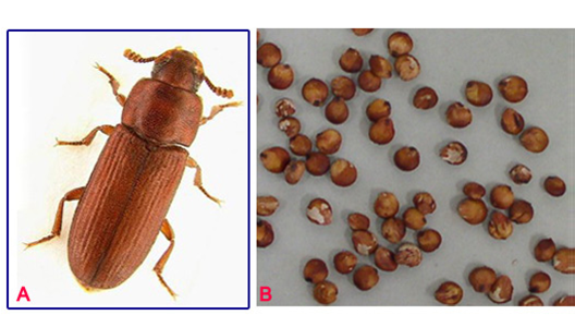 Red Flour Beetle (Tribolium castaneum) of pearl millet: (A)adult and (B)damaged grains (photos: ICRISAT). |
Hosts
Arachis hypogaea (groundnut), Avena sativa (oats), Bertholletia excelsa (Brazil nut), Hordeum vulgare (barley), Juglans (walnuts), Culinaris (lentil), Oryza sativa (rice), Phaseolus (beans), Phaseolus lunatus (lima bean), Pisum sativum (pea), Prunus dulcis (almond), Secale cereale (rye), Triticum (wheat), Triticum spelta (spelt) and Zea mays (maize).
Geographic distribution
T. castaneum is cosmopolitan in distribution, found in infesting stored grain, seeds, flour, dried fruits and nuts (Teetes et al. 1983).
Biology and transmission
The adult is 2.3-4.4 mm long, rather flat, oblong and chestnut-brown (reddish-brown). It lays about 450 eggs in stored grain. The eggs are minute, cylindrical and white. The incubation period lasts for 5-12 days. The yellowish-white cylindrical grub is covered with fine hairs. The pupa is naked (without a cocoon), yellowish-white, becoming brown later and adults emerge in 3-7 days (Teetes et al. 1983).
Detection/indexing methods at ICRISAT
- X-ray radiography is used for suspected samples because it offers a non-destructive test of seed samples. Dry seed examination using magnifying lens to separate the infested seed.
Treatment/control
- Fumigation of the samples with methyl bromide by 32 g/m3 for 4 hr followed by seed treatment with chlorpyriphos at 3 g/kg-1 seed (Ghanekar et al. 1996).
Procedures followed in case of positive test at ICRISAT
- Rejection of infested seed samples in case of positive test.
References and further reading
Chakrabarty SK, Anitha K, Girish AG, Sarath Babu B, Prasada Rao RDVJ, Varaprasad KS, Khetarpal RK, Thakur RP. 2005. Germplasm exchange and quarantine of ICRISAT mandate crops. Information Bulletin No. 69. Rajendranagar 500 030, Andhra Pradesh, India: National Bureau of Plant Genetic Resources; and Patancheru 502 324, Andhra Pradesh, India: International Crops Research Institute for the Semi Arid Tropics. 80pp.
Ghanekar AM, Ranga Rao GV, Murthy KS, Surender A, Shaik Babu Saheb. 1996. Seed protectants for healthy exports. Indian Journal of Plant Protection 24: 37-43.
Teetes GL, Seshu Reddy KV, Leuschner K, House LR. 1983. Sorghum insect identification handbook. Information bulletin No.12. Patancheru, A.P., India: International Crops Research Institute for Semi-Arid Tropics. 125pp.
Guidelines for the safe transfer of pearl millet germplasm
Contributors to this page: ICRISAT, Patancheru, India (RP Thakur, AG Girish, VP Rao).
Technical Guidelines for the Safe Transfer of Germplasm and the Protection of CGIAR Germplasm Banks
Pathogens of quarantine significance of pearl millet tested by the Germplasm Health Laboratory of ICRISAT
Pearl millet [Pennisetum glaucum (L.) R. Br.]
|
Bacteria
|
|
Pseudomonas syringae pv. syringae van Hall.
|
|
Xanthomonas campestris (Pammel) Dowson
|
|
Xanthomonas vasicola pv. holcicola (Elliott).
|
|
Fungi
|
|
Bipolaris setariae (Saw.) Shoem.
|
|
Claviceps fusiformis Loveless
|
|
Moesziomyces penicillariae (Bref.) Vanky.
|
|
Pyricularia grisea (Cke.) Sacc.
|
|
Sclerospora graminicola (Sacc.) Schroet.
|
|
Insects
|
|
Plodia interpunctella Hubner.
|
|
Prostephanus truncatus Horn.
|
|
Sitotroga cerealella Olivier
|
|
Tribolium castaneum Herbst.
|